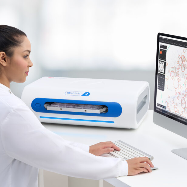
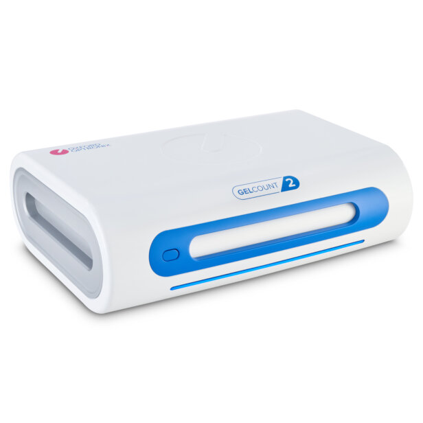
What is GelCount?
GelCount is an integrated hardware and software platform that images multi-well plates or Petri dishes containing cultured mammalian cell colonies, spheroids or organoids and processes the images obtained to yield a per well or per dish count of objects detected and object diameter statistics, all of which can be exported directly into Excel.
What’s unique about GelCount?
GelCount is conceived specifically for objective and reproducible detection and analysis of non-adherent mammalian cell colonies, spheroids or organoids in semi-solid media, an otherwise painstaking procedure carried out under a microscope.
How does GelCount work?
GelCount combines high-resolution / high depth-of-field CCD scanning hardware with a software-controlled sample loading mechanism that automatically sends images to a PC via a USB bus. Image processing occurs via proprietary software residing on the PC incorporating a powerful, purpose-written image processing algorithm.
What are the applications for GelCount?
GelCount will be of interest to cancer biologists utilizing colony, spheroid or organoid formation to measure the therapeutic response of drug candidates and other treatment regimens, typically on cancer cells in vitro. Secondary applications include the cell proliferation assay, the invasion assay or bacterial and yeast colony counting.
Is GelCount suited to counting adherent, stained colonies?
Yes! The GelCount is also perfectly suited to imaging and accurately detecting stained, adherent cell colonies in multi-well plates and Petri dishes.
Do colonies, spheroids or organoids in 3D media need to be stained?
Generally no. In most circumstances GelCount will reliably detect colonies, spheroids or organoids cultured in 3D assays (soft agar, Matrigel, etc.) without the need for cell staining. In some instances (e.g. high background, or very small spheroids/organoids) performance can be enhanced with the use of MTT-based metabolic stains. These do not stain the medium, just live cells and cell aggregates (recommended protocols available on request).
Can GelCount be used to count bacterial and/or yeast cell colonies?
Yes! While not a primary application, GelCount can be used very successfully to image and count bacterial or yeast colonies on standard, non-opaque agar types (example images available on request).
Is GelCount a cell counter?
Strictly speaking no. GelCount does not use magnifying optics and was conceived to detect cell aggregates (colonies, spheroids, etc.) of approximately 30 cells or more, rather than individual cells. However, at its maximum resolution GelCount is capable of distinguishing individual adherent cells, provided they are well stained and discrete. Under ideal conditions therefore, GelCount may be useful for quantifying the output of cell proliferation assays (example images available on request).
Does GelCount support inclusion/exclusion of colonies, spheroids or organoids based on size?
Yes, the image analysis algorithm offers extensive user-optimization, including minimum and maximum object diameter thresholds. User-generated detection settings can be stored for future analysis sessions.
What is the minimum object size GelCount can detect?
Depending on the morphology of colonies, spheroids or organoids being imaged, the spacing between them, and the overall contrast conditions, GelCount can detect objects as small as 50 µm in diameter at the maximum available imaging resolution setting.
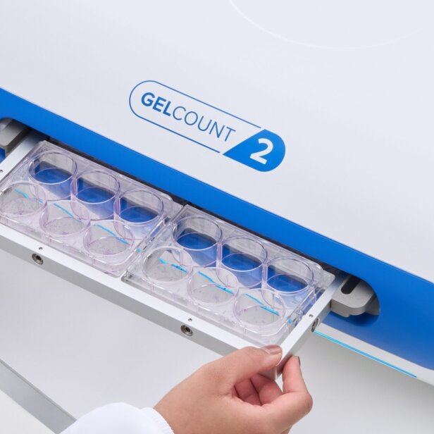
What plasticware types are supported?
GelCount is compatible with multi-well plates from 6- to 96-well plates, as well as with 35 mm, 50 mm, and 100 mm Petri dishes.
How fast is GelCount and what throughput rates can be achieved?
The rate limiting step is image acquisition, which is a function of the chosen imaging resolution and of the plate/dish type in use. The GelCount will require approximately 5 minutes to image and process four 6-well plates at 600 dpi (a typical case), or under 30 minutes to image and process four 96-well plates at maximum resolution (a demanding case).
Can GelCount analyse multiple samples simultaneously?
Yes! Up to four multi-well plates, up to four 100 mm Petri dishes, up to twelve 50 mm Petri dishes, or up to twenty-four 35 mm Petri dishes may be loaded, imaged and processed simultaneously.
Is GelCount compatible with a plate stacker or robotic loading system?
At the present time GelCount does not support ‘off-the-shelf’ stacker or robotic sample loading solutions.
Does GelCount support Mac OS?
No, GelCount software is currently only available for PC / Windows®.
What are the recommended PC specifications for GelCount?
Multi-core processor, 16 GB system memory or above, solid state storage, USB 2.0 or 3.0, Windows 11, Microsoft Excel pre-installed.
Can I test the GelCount?
Yes, depending on geographical location we are able to offer an on-site demonstration or short term product evaluation. A one-to-one screenshare-based product and software introduction is also offered. Please contact our sales team to explore these options.