Executive Summary
The Colony Forming Unit (CFU) or Colony Forming Cell (CFC) assay is universally recognised as the gold standard method for measuring the effect of radiation, chemotherapeutic drugs and other agents on cell viability. However, manually counting the resulting cell colonies is a laborious task in which consistent objectivity is difficult to achieve.
Commercial and funding pressures, resource limitations and the growing complexity of CFC assays means cancer biologists need to maximise colony sample processing capabilities while maintaining the rigour of standardised and accurate counts.
Automating the detection, counting and analysis of mammalian cell colonies offers significant benefits to cancer biologists processing tumour colony forming assays – eliminating the risk of subjectivity, bias and human error, increasing speed and accuracy, and delivering unprecedented data archiving and retrieval capabilities.
With its dramatic effect on throughput, automated colony counting opens up new possibilities for ambitious new projects that will advance the frontiers of research – including large-scale combinational studies.
Furthermore, today’s intelligence-based imaging systems for automated colony counting enable the collection, processing and archiving of ‘digital samples’ - adding a powerful new dimension to the peer review and reporting process. They also deliver additional quantitative data and make innovative collaborative research and validation approaches possible.
Introduction
The financial crisis of the past few years has led to deep cuts in public and non-government funding in the field of cancer research, resulting in hiring freezes across academic institutes. In 2012 Cancer Research UK, one of the largest charities in the UK, announced a 10% cut in basic research funding until 2015 because of a dip in income, which has had an impact on the pace of research and discovery.
It’s a similar story in the life sciences industry and the world of ‘big pharma’, where burgeoning development costs and increased regulatory scrutiny have caused R&D costs to spiral.
Against a backdrop of financial and resource constraint, today’s commercial and academic research laboratories are under pressure to perform. Delivering output that meets, in the case of commercial labs, stringent reporting requirements set down by healthcare regulators, while publicly funded medical research programmes must submit their findings to world-class peer review.
The resource squeeze has resulted in a re-evaluation of the drug discovery approach. One outcome has been a growing consensus that new biopharmaceutical models are needed to maximise investment outcomes relating to the development of new drugs and treatment regimes.
This has led to the emergence of ‘open innovation’ approaches to R&D and drug discovery, where the collective expertise of a network of contributors – academic researchers, commercial research firms, external investigators, service providers and biotechnology companies – is harnessed and the submission of promising assays encouraged.
In Europe, the drive is on to improve knowledge transfer and stimulate research collaborations between public research centres and industry. For example, in 2012 the European Commission issued voluntary operational guidelines for universities and other research institutions to improve links with industry and implement open access measures that make research outputs more readily available, while national bodies in the UK and Germany are in pursuit of similar knowledge sharing frameworks.
Clearly, participating and performing in this brave new world means today’s research laboratories need to be able to maximise output and throughput, demonstrate research granularity, and be equipped to participate in large scale (international) studies that may include multiple internal or external stakeholders.
1. Counting the cost
The issues and challenges of the colony formation assay technique
The colony formation assay is the established gold standard for measuring the effects of cytotoxic agents on cancer cells in vitro and is frequently used in cancer research laboratories to determine the effects of drugs or radiation on proliferating tumour cells. The treatment is usually a drug, ionizing radiation exposure or a combination of the two.
Error prone and resource intensive
To maintain consistency in data acquisition, utilising colony-based assays often requires that a single observer count colonies in hundreds, potentially thousands of
cultures. Clearly, the cost and time implications of this labour intensive approach are significant.
Observers need to be well trained in colony detection and scoring, and expert at eliminating ‘background noise’. Meanwhile, observer fatigue means both accuracy and
reproducibility of quantification suffer severely.
When multiple observers are employed, observer fatigue is reduced but the accuracy and reproducibility of colony enumeration is still compromised due to observer bias and significant inter/intra observer variability.
The rise of 3D arrays
The manual counting scenario is challenging enough in adherent mono-layer ‘two dimensional’ samples. But in recent years sphere formation (or tumour sphere) assays, in which cells are seeded in a semi-solid matrix (gel) and which more realistically simulate the tissue state, have become increasingly popular with cancer biologists.
In these three dimensional (3D) in vitro culture systems, accurate visual recognition requires the use of a microscope, making consistent manual counting dramatically more labour intensive.
The increasing emphasis on combination treatment for cancers and the number of agents under development, plus the growing requirement for quantitative 3D assays that fully reflect drug/treatment effects, means research laboratories are under pressure to undertake ever more complex assays.
And finally, the variability factor
As well as being labour intensive and subjective, manual colony counting utilising a microscope has been shown to result in a significant increase in the degree of intra and inter-observer variability.
Colony forming assays – the drawbacks
The introduction of new advanced assays has highlighted a number of drawbacks relating to the application of colony counting:
- Assays require manual enumeration and are therefore highly subjective
- The assay is operator intensive, time consuming and expensive to perform
- High coefficients of variation (CV) can result
- The assay lacks external standards and controls that do not allow assay calibration or standardisation, making validation difficult
2. The way forward
Automated colony counting and image analysis
Today’s modern automated colony counting and image analysis systems offer an efficient and cost effective alternative to manual counting, delivering a significant reduction in turnaround time resulting from high sample throughput.
The automated image acquisition and analysis approach to colony counting has been validated and found to provide superior accuracy and precision compared to manual observer counting, reducing both intraand inter-observer variation. Using high depth of field imaging combined with powerful algorithms, unsurpassed colony detection performance includes the resolution of overlapping colonies and differentiation of real colonies from debris or other artefacts.
Furthermore, today’s intelligent systems enable operators to set defined colony thresholds for counts – for example, the exclusion of colonies based on size or colony shape-related parameters as well as general object detection sensitivity – and allow repetitive plate counting of non-adherent colonies
without staining cells.
Providing a standardized method for automated CFU analysis, research labs are able to reduce the cost of colony counting by increasing throughput and reducing work flow demands while increasing the consistency and accuracy of results.
Generating new data sets
Technological advancements have made it possible to incorporate extensive analysis parameters into today’s single instrument automated platforms. Giving research laboratories unique new insights and the opportunity to capture and report additional qualitative and quantitative data.
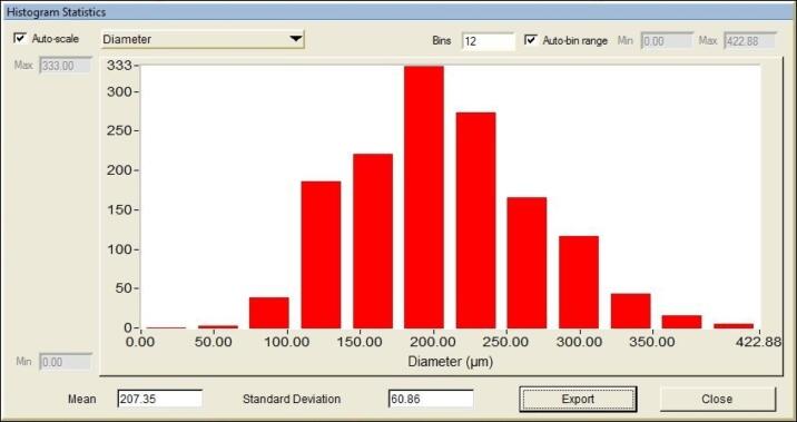
Researchers processing colony samples can now generate a count but also collate detailed colony size information in the form of a mean per well/dish, histogram distribution, or even on an individual colony basis.
This new found ability to quantitatively measure the effects of anti-cancer therapeutic regimes on absolute colony numbers and colony size makes it possible for research laboratories to extend the sensitivity of the colony forming assay, obtaining previously ‘hidden’ information relating to colony growth dynamics.
Data capture and export
Today’s sophisticated intelligence-based imaging systems for automated colony counting deliver unparalleled data capture and export capabilities.
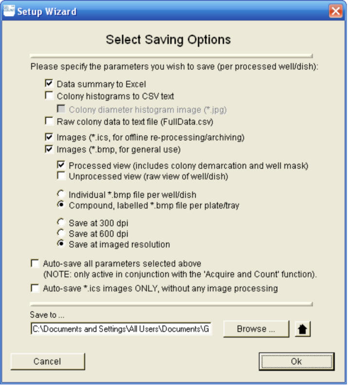
These extend from basic colony counts and mean colony statistics to full digital image archiving and per colony raw data export. Images of colony plates or dishes can be saved as a general-purpose bitmap format for presentations or print output into lab documentation, or in a digital raw image format that supports the future processing or reprocessing of samples.
Now research laboratories can visually document findings quickly or submit captured images to multiple research teams or external agents for independent validation and/or processing and assessment.
This ability to capture, process, export and data on or off line gives laboratories a cost effective and fast method of undertaking objective and rigorous double blind tests and extends the ability of laboratories to participate in global or large scale trials or pursue new collaborative research approaches.
Reproducible assays
Finally, the capabilities of laboratories to engage in large scale or multi-site research projects are further extended by the ability to store and reapply defined count parameters, to maximise counting proportionality and reproducibility.
Settings can be stored and reutilised in single studies, reducing the complexity of accurately assessing the effects of drugs or combined therapeutic approaches in respect to both dose and time of exposure using fewer culture plates.
Count parameters and settings can also be stored and submitted to other teams or personnel, enabling in parallel validation programmes or the multiplication of treatment assessment trials.
The benefits of automated colony counting
- Rapid, objective and statistically significant
- Not subject to bias
- Capable of processing high volumes of samples in a standardised and consistent manner
- Enables accurate, single-pass detection of colonies in 3D assays
- Enables reliable and reproducible counts
3. An integrated approach: GelCount™
GelCount is an easy-to-use PC software operated colony counter that automates the detection, counting and analysis of mammalian cell colonies in Petri dishes, flasks and multi-well plates.
The first, and only, imaging system to enable objective and reproducible colony detection, specifically conceived for the cancer biologist, GelCount delivers unprecedented data archiving and retrieval capabilities that make it possible for laboratories to engage in ambitious new projects and collaborative approaches – including large-scale combinational studies and multi-site projects – and submit findings for peer review and verification.
Providing a powerful and cost-effective alternative to the labour-intensive task of manual colony counting, colonies are imaged, transferred, processed and characterised. The data is collated and exported within a single integrated hardware/software platform, eliminating the need to image samples on one device and processes these within an independent image analysis package.
This integrated approach enables:
- Images to be captured and processed within a single integrated hardware/software platform
- Instant archiving and output of raw colony images
- Off-line processing/reprocessing – even when original samples are no longer available
- Detection parameters (colony size/shape) can be previewed in real-time and stored for reuse
- Data findings can be captured and submitted for independent review on workstations where GelCount software is installed
- Data can be analysed and exported in a variety of formats.
Impressive performance
GelCount provides an effective and reliable resolution to the problem of counting colonies manually, delivering a dramatic effect on throughput while eliminating the impact of human error that results from subjective interpretation, bias or fatigue.
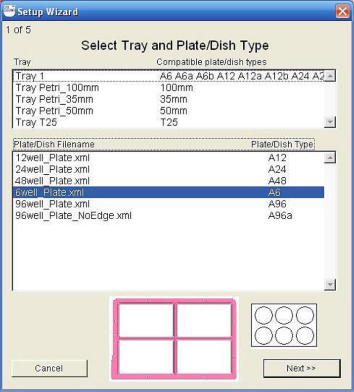
Compatible with multi-well plates (6-, 12-, 24-, 48- and 96-well), with Petri dishes (35mm, 50mm and 100mm) and with T25 flasks, GelCount’s high depth-of-field imaging allows the single-pass detection of colonies as small as 30 µm in diameter in medium layers of up to 5mm in depth.
GelCount delivers impressive sample throughput and colony processing times (from image acquisition to analysis and data exportation):
- Adherent colonies – typically 5 or 6 minutes per four 6-well plates
- Non-adherent colonies (in soft agar or equivalent) – typically 12-15 minutes per four 6-well plates
Powerful parameter setting
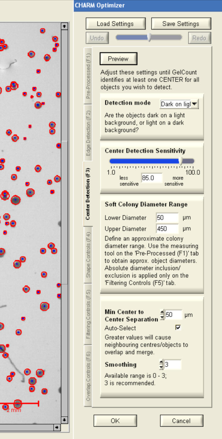
Colony detection is fully user-definable with the ‘CHARM Optimizer’ utility, which allows the adjustment of numerous sensitivity and shaperelated parameters, including the crucial ability to include or exclude colonies based on size.
Adjustments can be pre-viewed in real-time and parameters stored as templates for reuse in future experiments.
Objective and reproducible detection and analysis
GelCount enables researchers to go from colony samples to colony counts, collecting additional statistical data such as colony size information at the click of a button.
- Objective application of user-definable colony detection rules, including colony size thresholds – user settings can be stored and instantly reapplied, or distributed to remote colleagues and collaboration partners
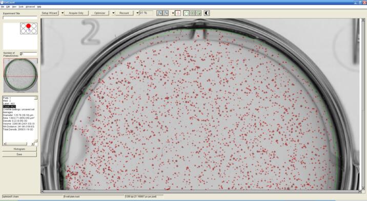
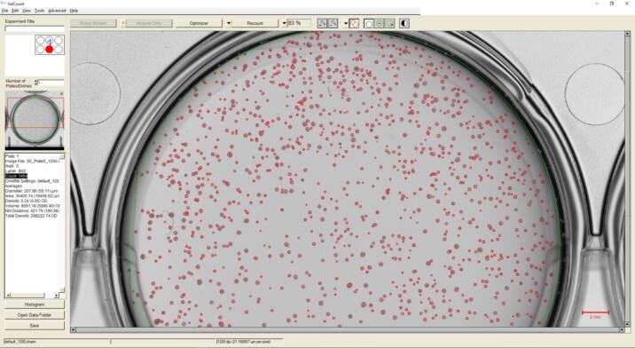
- Impressive colony detection capabilities; resolution of overlapping colonies, differentiation of real colonies from debris, suppression of false positives resulting from contamination or staining/agar artefacts (see image)
- Supports counting and analysis of less discrete colonies – even in 3D assays.
Instant data capture and data export
Images of colony samples are automatically captured and transferred to a PC for archiving. These images can be output (printed) or utilised in bitmap format for digital presentations/submissions. An optional raw image type also supports the processing or reprocessing of images ‘off-line’ from any independent workstation on which GelCount software is installed.
GelCount’s data management and exportation capabilities are extensive:
- Generation of histograms containing a unique set of statistical distributions, such as colony size; histogram data can be manually or automatically exported in bitmap or Excel® compatible raw data formats
- Automated exportation to Excel® of summary data, including colony counts and statistics
GelCount: Key Features
Integrated and dedicated colony counting platform
All-in-one solution for image acquisition and image processing.
No microscope required
High-resolution, high depth-of-field, single-pass detection of colonies of 30 μm diameter or above.
Versatility
Adherent or non-adherent cell/colonies types; compatibility with multi-well plates, Petri dishes and T25 flasks.
Objective and consistent
‘CHARM’ algorithm objectively and consistently applies user definable colony detection parameters.
Performance / throughput
Typically <15 minutes or high-resolution imaging and processing of four 6-well plates.
Colony sizing
Automatically generates colony diameter information and other statistical parameters.
Excel® and image exportation
Direct exportation of counts and summary data to Excel®; raw or generic bitmap image exportation
User-friendly
Sophisticated image analysis software with an intuitive and efficient user interface.
Generous feature package
Unlimited software licenses; unlimited software updates; 2-year product warranty; free product support
You can review a more detailed GelCount™ feature description here.
4. GelCount™ in action
Compared to manual counting, GelCount delivers significant performance enhancements including faster processing and throughput, improved data granularity and accuracy – all of which saves valuable laboratory time without compromising quality and opens the way for researchers to apply improved preclinical methodologies to study drug effects on cancers.
For example, by reducing the labour and time investment involved in the analysis and validation of experiment outcomes against defined oncology targets, GelCount enables even small scale research laboratories to cost effectively undertake large scale or complex combinational studies.
Extending the boundaries
In a 2008 study A new preclinical 3-dimensional agarose colony forming assay*, researchers at the University of Texas outlined how GelCount enabled the precise study of drug effects and made it possible for them to establish a reproducible and easy-to-perform quantitative 3D assay that fully reflected drug action on both colony size and number.
In their paper, the researchers document how GelCount’s technology made it possible to implement a new 6- or 12-well soft agar assay, telescope analysis timeframes and undertake:
- repetitive plate counting over time and under different conditions, without staining cells
- monitoring of the effects of drug treatment over time
- identification of differentials in colony formation
- undertake image capture of drug effect on colony size and number – supporting the fast identification of new drug combinations
- cell agnostic counts –researchers were able to implement a range of five cell lines, including bothgliomas and adenocarcinomas
* Kajiwara Y, Panchabhai S and Levin V (2008) A new preclinical 3-dimensional agarose colony forming assay, Technology in Cancer Research and Treatment, Vol 7 (4).
“We have now taken this best practice of industrialized colony counting and purchased the [GelCount] for our drug discovery activities.”
Dr. Jannik N Anderson, Institute for Applied Cancer Science, University of Texas
“GelCount reduces the analysis time considerably when compared to manual counting, and the results are similar between them. It has also the added bonus of reducing the subjectivity that could arise by manual counting, making the clonogenics more reliable.”
Dr. Natividad Gomes-Roman, Institute of Cancer Sciences, University of Glasgow, Beatson Institute for Cancer Research, Glasgow, UK
“I can easily image all the wells, and after choosing my desired settings, in a short time I can have the number of my colonies and their size. GelCount allows me to follow the growing of the colonies in time by simply acquiring different images during the weeks, so that I can see the effects of the different sample treatments. Definitely, the use of the GelCount saves a lot of my time and now I can perform more experiments in a shorter time, with the additional advantage of acquiring a complete and clear image of the entire well for presentation purposes.”
Dr. Tiziana Scanu, The Netherlands
5. Concluding thoughts and future opportunities
Recent years have seen an explosion of new discoveries in relation to the diverse molecular and biological changes underlying cancer development and progression. These insights are changing our understanding of the complex pathways that regulate cancer cell biology and the mechanisms that restrain tumorigenesis. Researchers are translating these findings into novel approaches towards cancer diagnosis, prognosis and therapies.
However, such developments are generating some fundamental operational challenges for cancer research laboratories. The expanding number of treatment drug/therapy options and trial candidates means CFC arrays are mounting in scale and complexity as researchers attempt to evaluate increasing numbers of variables.
The emergence of 3D arrays and the ongoing advances in array development means today’s research laboratories need the capacity to undertake ‘industrial scale’ assays. However, the time intensive and laborious manual colony counting approach may prove too cost inhibitive for some laboratories and ultimately may preclude them from participating in future cutting edge research.
The economic downturn has significantly impacted public and non-governmental research funding. Today’s academic research laboratories are under pressure to firstly secure funding and then maximise grant spend to undertake research.
It’s a similar story for commercial and contract research companies, where the pressures are mounting to maximise throughput and utilisation of facilities and resources, including highly qualified research technicians.
Universally, research labs from all sectors – private, public and academic – need to demonstrate research rigour, accurately report research findings, and engage in collaborative or peer review programmes. Automated colony counting and imaging make it possible to deliver against all these requirements.
Employing state-of-the-art image acquisition, processing and analysis platforms delivers an accurate, precise and automated analysis system. For the future, improvements on the automation horizon – such as the robotic loading of samples and increased sensitivity of data collection parameters – are set to deliver deeper, more accurate data analysis.
All of which opens up exciting new possibilities that will advance the frontiers of cancer research.
To view GelCount’s full citation list, here.