Introduction
Acute kidney injury, in a simple definition, is when kidneys abruptly stop functioning and cleansing the blood of its waste and toxins. Within hospitals, this outcome directly leads to a higher probability of patients having much longer stays in or, in the worst case, dying in hospital, both of which drastically increases healthcare costs. Acute kidney injury occurs in approximately 30% of patients after heart surgery and within another 50% of patients who acquire sepsis after surgery, which demonstrates how large of an issue this is for hospitals and staff.
Despite this condition being so prevalent, there are currently no treatments or therapies to prevent kidney injury after pulmonary surgery or sepsis. This is primarily due to a poor understanding of its causes, and the inability of doctors to assess the risk of developing kidney injury before it shows deadly symptoms.
A new approach to a common problem
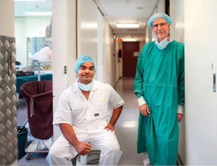
This lack of understanding has led to an experienced team of researchers and clinicians from the Florey Institute of Neuroscience and Mental Health to examine ways to reduce the prevalence of renal injury following heart surgery and sepsis. By using clinically relevant sheep models, Dr. Yugeesh Lankadeva and colleagues have revealed that reduced oxygen and microvascular blood flow levels in the renal medulla (the inner part of the kidney) is a frequent pathological feature after heart surgery and during sepsis, which may be driving acute kidney injury.
This group has further demonstrated that it is possible to reliably estimate oxygen levels within the kidney’s by assessing oxygen within bladder urine. The placement of a catheter into the bladder is standard clinical practice in patients undergoing pulmonary surgery in operating theatres and patients treated for sepsis in intensive care units. Therefore, observing bladder urine oxygen levels in these patients is a relatively non-invasive procedure for the early detection of those at risk of developing acute kidney injury. Dr Lankadeva’s recent findings also suggest that it could be feasible to alter existing therapeutic strategies to enhance microvascular blood flow and oxygen levels within the renal medulla. This may decrease the prevalence of acute kidney injury arising from heart surgery and sepsis.
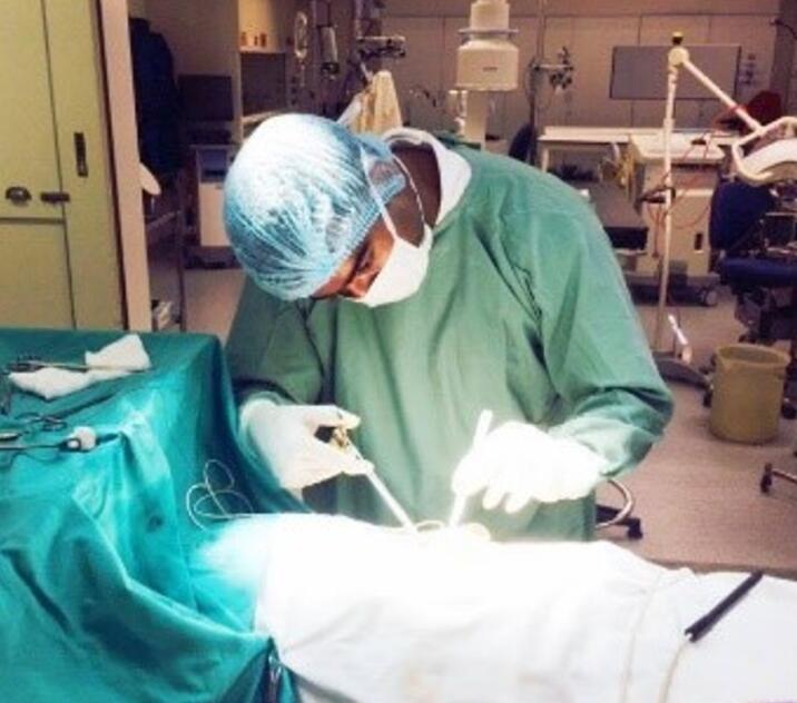
The need for effective therapeutic strategies for acute kidney injury is persuasive, as it will lower the short-term risk of death. Moreover, effective strategies will also lower the long-term risk of chronic kidney disease, end-stage renal disease and cardiovascular diseases. The development of techniques by Dr. Lankadeva and colleagues to simultaneously monitor microvascular tissue perfusion using OxyFlo™ Pro and oxygen levels using OxyLite™ Pro in both anaesthetized and non-anaesthetized animals presented these researchers a highly powerful and unique tool to explore the causes of organ failure arising from heart surgery and sepsis.
Highlight publications
Ngo JP, Lankadeva YR, Zhu MZ, Martin A, Kanki M, Cochrane AD, Smith JA, Thrift AG, May CN, Evans RG. Factors that confound the prediction of renal medullary oxygenation and risk of acute kidney injury from measurement of bladder urine oxygen tension. Acta Physiol; 2019, 227: e13294
Lankadeva YR, Cochrane AD, Marino B, Iguchi N, Hood SG, Bellomo R, May CN, Evans RG.
Strategies that improve renal medullary oxygenation during experimental cardiopulmonary bypass may mitigate postoperative acute kidney injury. Kidney International; 2019, 95:1338-1346
Iguchi N, Kosaka J, Booth LC, Iguchi Y, Evans RG, Bellomo R, May CN, LankadevaYR.
Renal perfusion, oxygenation, and sympathetic nerve activity during volatile or intravenous general anaesthesia in sheep. British Journal of Anaesthesia, 2019 122, 342-349
LankadevaYR, Kosaka J, Iguchi N, Evans RG, Booth LC, Bellomo R, May CN. Effects of Fluid Bolus Therapy on Renal Perfusion, Oxygenation, and Function in Early Experimental Septic Kidney Injury. Critical Care Medicine, 2019, 47 (1): e36-e43
Lankadeva YR, Kosaka J, Evans RG, Bellomo R, May CN. Urinary oxygenation as a surrogate measure of medullary oxygenation during angiotensin II therapy in septic acute kidney injury. Critical Care Medicine, 2018, 46 (1): e41-e48
Lankadeva YR, Kosaka J, Evans RG, Bailey SR, Bellomo R, May CN. Intra-renal and urinary oxygenation during norepinephrine resuscitation in ovine septic acute kidney injury. Kidney International, 2016, 90 (1): 100-108
Author: Justin Croft, March 2020