Introduction
Scaffolds are support structures used in tissue engineering that are designed to assist cellular growth and proliferation of certain cells upon implantation onto a specific tissue. A fundamental property of scaffolds is their biocompatibility. In its most general sense, biocompatibility may be characterized as the ability of a biomaterial to perform its desired function with respect to a medical therapy, without eliciting any undesirable local or systemic effects in the recipient or beneficiary of the therapy. As such, these biomaterials are most often developed in vitro first and then fully fleshed out within animal models.
As biocompatibility is important, researchers do attempt to make their cellular models and scaffolds work as close to what would be seen in the in vivo condition. As oxygen is one of the most important molecules for sustaining life, it is understood that it is also an important variable to consider in tissue engineering and regenerative medicine. It has been shown that the change in oxygen concentration in an artificial or tissue-engineered graft affects cell survival, differentiation, and tissue growth in profound ways. As researchers continue to need to control and identify oxygen levels within scaffolds, Oxford Optronix is in a unique position to help these researchers on both ends of this spectrum.
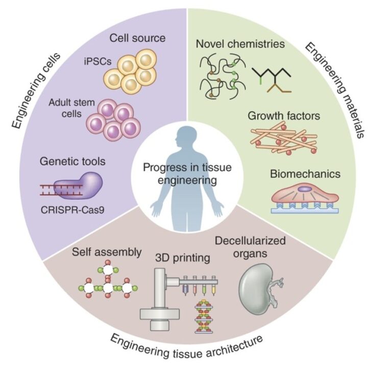
The image is a summary of the 3 primary tissue engineering areas, namely engineering cells (stem cells and genetic tools), engineering materials (growth factors, chemistries, and biomechanics), and the available tissue architectures (self-assemblies, 3D printing, and decellularized scaffolds).
In vitro scaffold research and Oxford Optronix
For in vitro studies, cells grow around and within scaffolds and are heavily dependent on the external and internal oxygen environment. Here, researchers can test scaffolds in media within hypoxia chambers (such as the HypoxyLab) or within bioreactors that control the flow of physoxic media over a scaffold. Within these closed systems researchers can measure the pO2 both within their media and their scaffolds, using the small OxyLite oxygen sensor.
References
Below are some recent publications in this area.
Maury et.al. 2021
Rapid Evaluation of Novel Therapeutic Strategies Using a 3D Collagen-Based Tissue-Like Model
Used to HypoxyLab to set O2 levels of the 3D to cell collagen-based models. Then used the OxyLite monitor to measure the collagen models pO2 levels in real time.
Niu et. al. 2020
High Oxygen Preservation Hydrogels to Augment Cell Survival under Hypoxic Conditions
Oxygen tension in the hydrogels with or without MSC encapsulation was measured using an OxyLite fibre-optic oxygen micro-sensor.
Lee et. al. 2017
Developing a Customized Perfusion Bioreactor Prototype with Controlled Positional Variability in Oxygen Partial Pressure for Bone and Cartilage Tissue Engineering
Used the OxyLite sensors to take pO2 readings across the tissue cultivation platform within a closed bioreactor.
Nyga et. al. 2013
A Novel Tissue Engineered Three-Dimensional in vitro Colorectal Cancer Model
Oxygen levels were measured in 3-D constructs comprising the artificial cancer mass using the OxyLite tissue oxygenation monitoring system.
Download this article
Author: Justin Croft, February 2022