Introduction
A clean incubator is essential for reliable cell-culture and microbiology results. Even low-level contamination can add variability and force costly repeats. This protocol covers cleaning frequency, approved agents, a deep-clean workflow, and how to use MycoFog™ between full cleans.
To clean a CO₂ incubator or hypoxia workstation, power down, remove all items, and wipe internal surfaces, shelves, and the door gasket with 70% ethanol or an approved disinfectant. Disinfect the water tray and allow everything to dry before reassembling. For added protection between experiments, a hydrogen peroxide fogging system such as MycoFog™ can be effective at reducing contamination risk further.
Note: this guide is intended for laboratory CO₂ incubators and hypoxic workstations used in research, not neonatal or NICU incubators, which require clinical-grade procedures.
Recommended cleaning schedule and frequency
Frequency depends on usage intensity and how critical contamination control is for your work.
- Daily / weekly wipe-down: Disinfect high-touch areas (handles, gasket, shelves). Shared or high-throughput labs should wipe daily with 70% ethanol; lower-use labs may manage with a weekly wipe. Routine wipes markedly reduce microbial load.
- Monthly deep cleaning: Shut the unit down and fully clean interiors, removable parts, and the humidity pan about once a month (every 2–3 months at minimum). Structured monthly cycles have been associated with large drops in contamination incidents.
- Immediate cleaning: Clean and disinfect after media spills, visible mould, cloudy reservoir water, unusual odours, or culture outbreaks. Prompt action, including a heat or MycoFog™ cycle, stops a small issue becoming a chamber-wide problem.
Keep a cleaning log and make sure all users know the schedule and their responsibilities.
Approved cleaning agents and supplies
Use agents that kill contaminants without damaging stainless steel, gaskets, or sensors. Always check the manufacturer’s manual first.
- 70% ethanol or isopropanol: Good for routine wipe-downs. Fast-evaporating and effective against many bacteria and fungi, though not spores or some non-enveloped viruses.
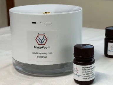
Hydrogen peroxide (3–8%): Broader activity, including better effect on viruses and spores. Safe for most interiors; breaks down to water and oxygen. Use on a cloth or as a fog via MycoFog™.
- Manufacturer-specific products: Preferred for deep cleans when available, because they are tested on incubator materials.
- Water-pan treatments: Add sterile copper sulphate, H₂O₂ tablets, or products such as AquaGuard® or Preventol® when changing water. Some systems (e.g. HypoxyLab) also use UV in the reservoir.
- Avoid: Bleach, strong corrosives, quaternary ammoniums, phenolics, and abrasive pads unless explicitly approved. These can corrode steel, damage sensors, leave residues, or scratch surfaces.
Important: unapproved chemicals can void warranties. Contact the supplier if uncertain.
Step-by-step cleaning procedure
Use this workflow for a monthly deep clean or after a contamination event. Gather gloves, disinfectant, sterile water, and PPE before starting.
Prepare — power down and empty
Turn off gas and power and unplug the unit. Remove cultures, racks, shelves, and trays (transfer cultures to a backup incubator if needed). Discard contaminated materials and old humidity-pan water as biohazard waste. Wear a lab coat, gloves, and eye protection.
Clean internal surfaces and components
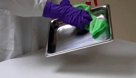
Wipe walls, floor, door interior, and ceiling with 70% ethanol or approved disinfectant, including corners and edges. Scrub removable shelves, brackets, and the water pan; rinse if soap was used. Clean fan blades and housings only when unplugged; wipe around sensors without soaking them. Replace non-cleanable HEPA filters on schedule. Thoroughly wipe gasket folds with swabs if needed.
Disinfect the water tray
Empty and scrub the humidity pan at least monthly to remove biofilm and scale. Rinse with sterile or distilled water and refill with fresh sterile distilled water plus an approved antimicrobial additive. Follow the manufacturer fill line. Consult the manual for automatic humidification systems.
Dry and reassemble
Air-dry all surfaces and parts completely. Inspect the gasket and fittings; replace corroded parts. Reinstall shelves level, refill the water tray, and secure sensors, fans, and filters.
Start-up and verify
Restore power and gas and wait for temperature, CO₂, O₂, and humidity to stabilize. Check calibration if required. Optionally run a manufacturer heat-sterilization cycle before returning cultures. Log the date, operator, and observations. Return only healthy, uncontaminated cultures
Integrating MycoFog™ into routine bio-decontamination
Manual wiping cannot reliably reach every crevice.
Hydrogen peroxide fogging with MycoFog™ complements deep cleans.
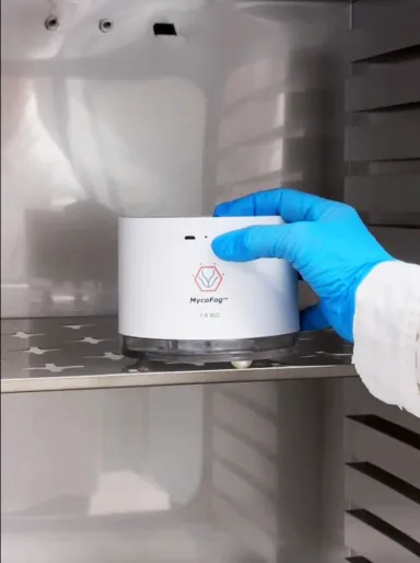
- Between deep cleans: Run a cycle weekly or biweekly (more often in shared or high-stakes incubators) without dismantling the chamber.
- After manual cleaning (optional): Fog the empty, dry, reassembled unit to treat missed crevices, then ventilate per the instructions.
- Quick turnaround: Fogging alone can reduce load after a scare until a full clean is possible. It disinfects surfaces; it does not replace removal of dirt or biofilm.
Effectiveness:
MycoFog™ mist has been validated for broad coverage and high-level disinfection, including 6-log kill of resistant bacterial spores and uniform vapor distribution into corners.
Safety:
The battery-powered unit runs a pre-programmed cycle with 7.8% H₂O₂ inside the closed chamber. Residual gas falls to safe levels by the end of the cycle; brief ventilation is typically enough and no post-wipe is usually required. Peroxide decomposes to water and oxygen.
Regular fogging is especially useful for multi-user incubators and sensitive cell lines: it resets surface bioburden with little labour or downtime.
Validation and monitoring
- Routine inspection: Watch for mold, residue, cloudy water, and recurring culture contamination.
- Swab testing: Periodic surface swabs (gasket, shelves) can confirm cleaning or highlight hotspots.
- Indicators: Chemical strips during fogging confirm vapor reach; biological indicators can verify kill where required (more common in GMP).
- Servicing: Annual or biannual manufacturer service checks sensors and seals. GLP/GMP labs may need formal re-qualification.
Conclusions
Physical cleaning remains essential; fogging adds chamber-wide decontamination without disrupting the schedule. Combined with approved disinfectants, documentation, and monitoring, this approach keeps incubators and hypoxia workstations a safe environment for cell cultures rather than a source of microbes.
For questions about incorporating MycoFog™ into your regimen, contact Oxford Optronix
References
- Abatenh E et al. (2018). Contamination in a Microbiological Laboratory. IJRSB
- Mizuno M et al. (2025). Cleaning methods for biosafety cabinet to eliminate residual mycoplasmas, viruses, and endotoxins after changeover. Regenerative Therapy
- Borges ED et al. (2019). Microbial contamination in assisted reproductive technology: source, prevalence, and cost. Journal of Assisted Reproduction and Genetics
- Meleties M et al. (2023). Vaporized Hydrogen Peroxide Sterilization in the Production of Protein Therapeutics: Journal of Pharmaceutical Sciences
- MycoFog (2024). MycoFog™ Efficacy & Safety Validation (internal validation report v2.5).
- MycoFog (2025). Use of Chemical Indicators to Validate the MycoFog® Biodecontamination System Cycle (White Paper). MycoFog Inc.